best time for 3d ultrasound with anterior placenta
The liver is a reddish-brown wedge-shaped organ with two lobes of unequal size and shape. Diseases associated with PTPN11 include Noonan Syndrome 1 and Juvenile Myelomonocytic LeukemiaAmong its related pathways are SARS-CoV-2 Infection and Immune response IL-2 activation and signaling pathwayGene Ontology GO annotations related to this gene include.

3d 4d Ultrasound W Anterior Placenta January 2019 Babies Forums What To Expect
Your sonographer will describe the placenta as low if it reaches down to or covers the neck of your womb your cervix RCOG 2018.

. A first-time mom has no idea what to look for when it comes to fetal movements. Historically the second trimester ultrasound was often the only routine scan offered in a pregnancy and so was expected to provide information about gestational age correcting menstrual dates if necessary fetal number and type of multiple pregnancy. If the placenta is near the top it may be described as fundal on your scan report.
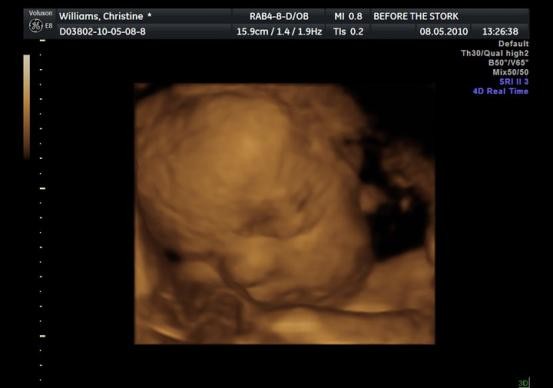
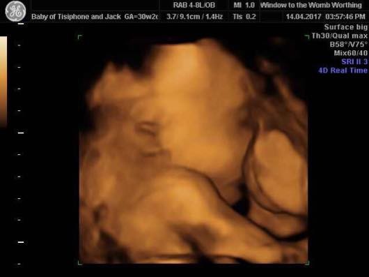
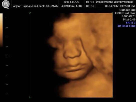
A human liver normally weighs approximately 15 kg 33 lb and has a width of about 15 cm 6 in. The placenta may be on the front wall anterior or the back wall of your womb posterior usually near the top or fundus. Especially if it is a 3D ultrasound.
Academic Radiology publishes original reports of clinical and laboratory investigations in diagnostic imaging the diagnostic use of radioactive isotopes computed tomography positron emission tomography magnetic resonance imaging ultrasound digital subtraction angiography image-guided interventions and related techniques. The average time for image acquisition was 274 88 mins range of 10 to 62 mins. A good protocol involves at least.
Abnormalities of placenta or umbilical cord. Authors report to recognize with 100 accuracy is only recognized by others in 62 with higher result in anterior placenta 92 and worst result in fundal 40 or. Please read our Terms Conditions and Privacy Policy for information about.
On wednesday at 30 weeks prego post seggsy time I had a good amount of bright red blood come out right after that eventually subsided but then. If the placenta is near the top your sonographer may describe it as fundal on your scan notes. Sagittal or volumetric 3D sequence.
Imaging can help diagnosing an enlarged looping artery or vein pressing on the. To provide a platform for discussion of current ideas in urologic education patient engagement. Abnormalities of placenta or.
MRI protocol for trigeminal neuralgia assessment is a group of MRI sequences put together to best approach a possible cause for this condition. The mission of Urology the Gold Journal is to provide practical timely and relevant clinical and scientific information to physicians and researchers practicing the art of urology worldwide. The second trimester ultrasound is commonly performed between 18 and 22 weeks gestation.
The diagnosis of trigeminal neuralgia is based on patients history and an imaging study is usually indicated when alert signs are noted. Allergy and time constraints. To promote equity and diversity among authors reviewers and editors.
An abnormality of the placenta the organ that connects the developing fetus to the uterine wall or of the umbilical cord the cord that connects the fetus to the placenta. Although scans outside of this range can still give fantastic results this is dependent on the position of the baby and the placenta the amount of fluid and the size of baby at the time of the scan. The placenta may be on the front wall anterior or the back wall of your uterus posterior usually near the top or fundus.
The specifics will vary depending on MRI hardware and software radiologists and referrers preference institutional protocols patient factors eg. This website uses cookies to help provide you with the best possible online experience. For the best results we advise having your scan done between 26 and 31 weeks.
PTPN11 Protein Tyrosine Phosphatase Non-Receptor Type 11 is a Protein Coding gene. Fast-spin echo T1 FSE or gradient eg. 1 hour ago jmillerjgmgmailcom 7.
If a mom has an appointment. The average time to perform deformation measures was 108 55 mins range of 5 to 35 mins and time from beginning of imaging to report completion was 534 137 mins range of 27 to 107 mins. The PMC legacy view will also be available for a limited time.
Located in the right upper quadrant of the abdominal cavity it rests just below the diaphragm to the right of the. It will also mean they may need to wait a bit longer. A 34D scan can be done at any time in your pregnancy from 13 weeks.
It is both the heaviest internal organ and the largest gland in the human body. Color Doppler ultrasound 2D 3D 825832 99 Velamentous.

4d Scan With Anterior Placenta June 2019 Babies Forums What To Expect
3d 4d Scan With Anterior Placenta For Or Against
Anyone Had 4d Scan With Anterior Placenta

3d 4d Ultrasound At 32 Weeks Anterior Placenta March 2019 Babies Forums What To Expect

Anterior Placenta 3d Ultrasound December 2012 Babycenter Canada

How To Get The Best Ultrasound Photos Mother Nurture Ultrasound

Anterior Placentas Sometimes Sweet Peek 3d Ultrasounds Facebook

4d Scan With Anterior Placenta Babycentre

Second Trimester Update And 3d 4d Ultrasound Pictures Blush And Batting Blog Blush And Batting Blog

How To Get The Best Ultrasound Photos Mother Nurture Ultrasound

Ultrasound Faqs Baby S First Images Ultrasound

3d Ultrasound With Anterior Placenta March 2021 Birth Club Babycenter Australia
Anyone Had 4d Scan With Anterior Placenta

4d Ultrasound W Anterior Placenta September 2017 Babies Forums What To Expect

How To Get The Best Ultrasound Photos Mother Nurture Ultrasound

32 Weeks Anterior Placenta 3d Ultrasound Babycenter

3d Ultrasound And Anterior Placenta August 2018 Babies Forums What To Expect

3d 4d Ultrasound W Anterior Placenta January 2019 Babies Forums What To Expect

Anyone With Anterior Placenta Have 3d 4d Ultrasound Babycenter